Sinus Tarsi Syndrome MRI Sumer's Radiology Blog
Sinus tarsi syndrome is the clinical syndrome of pain and tenderness of the lateral side of the hindfoot, between the ankle and the heel. Imaging often demonstrates the ligaments and soft tissues in the sinus tarsi are injured. Epidemiology
Sinus Tarsi Syndrome Radsource
The sinus tarsi (ST) is a small. MR imaging of the tarsal sinus and canal: normal anatomy, pathologic findings, and features of the sinus tarsi syndrome. Radiology. 1993; 186 (1):233-240. doi: 10.1148/radiology.186.1.8416571. [Google Scholar] 2. Stoller DW, Ferkel RD. The ankle and foot. In: Stoller DW, editor.

Sinus Tarsi Syndrome MRI Sumer's Radiology Blog
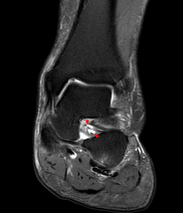
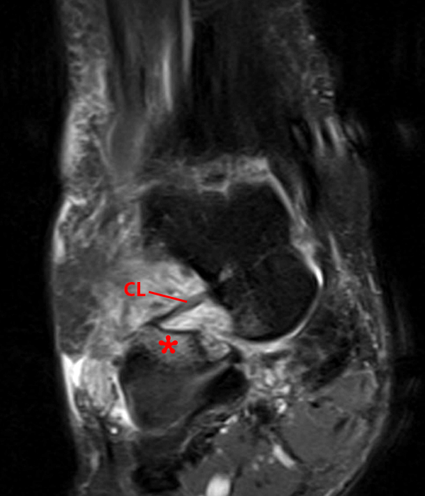
1. Introduction. The sinus tarsi is a cylindrical canal located in the hindfoot, bordered by the neck of the talus and anterosuperior calcaneus. The anatomy of this region is complex, with five ligamentous structures lying within it, including the intermediate, medial and lateral roots of the inferior extensor retinaculum, the cervical ligament (CL) and the interosseous talocalcaneal ligament.

Sinus tarsi syndrome Radiology Case
Journal of Radiology 80 (2011) e394-e400 Contents lists available at ScienceDirect European Journal of Radiology journa. anesthetics, has been described as the sinus tarsi syndrome [2,4,5]. The clinical syndrome can be attributed to trauma in about 70% and to chronic inflammation or deformity of the foot in the
Sinus Tarsi Syndrome Radsource
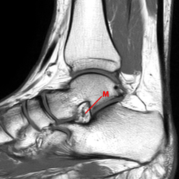
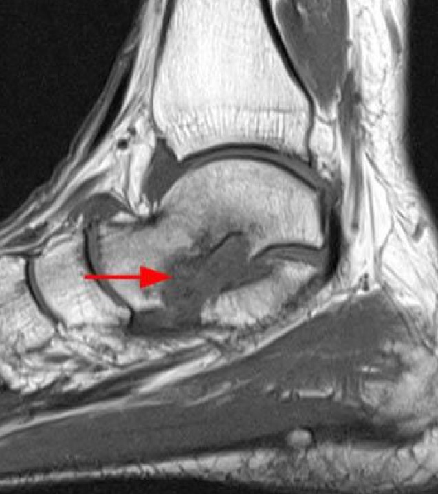
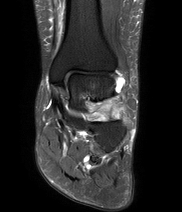
Abnormalities of the tarsal sinus and canal were seen on MR images in 33 cases (26.8%), were highly associated with tears of the lateral collateral ligament, and could be categorized according to the pathologic findings in patients with sinus tarsi syndrome: (a) diffuse infiltration with low T1- and T2-weighted signal intensity (n = 17) consiste.
Sinus Tarsi Syndrome Radsource
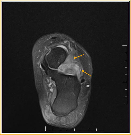
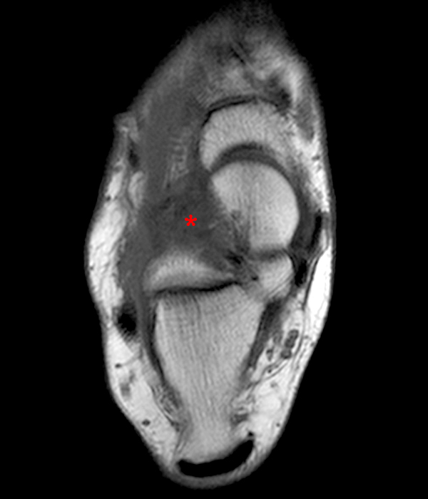
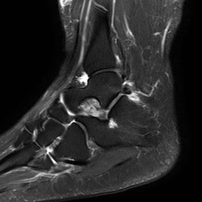
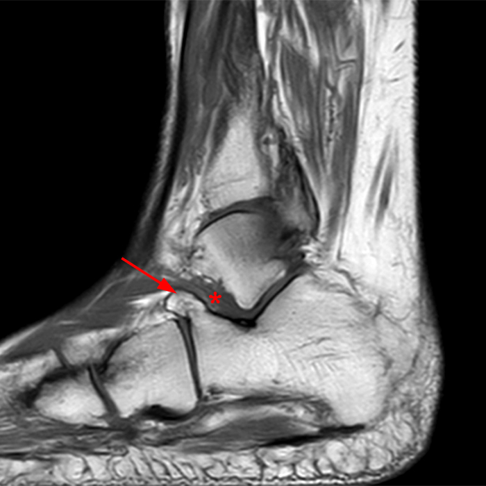
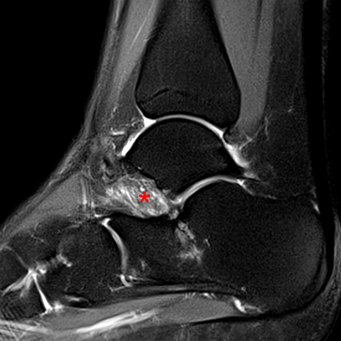
The sinus tarsi (ST) is a funnel-shaped region at the junction of the mid-foot and hind-foot containing fat, vessels, nerves and ligaments. The ligaments function to stabilize the subtalar joint and help maintain the longitudinal arch of the foot.

Sinus Tarsi Pain Explanation The Foot and Ankle Centre of Victoria
Sinus tarsi syndrome was first described in 1957 by Dr. Denis O'Connor [1]. The syndrome presents as pain in the lateral hindfoot, made worse following application of pressure to the opening of the sinus tarsi [2].. 2023, European Journal of Radiology. Show abstract. The sinus tarsi is a funnel-shaped region at the junction of mid-foot and.
Sinus Tarsi Syndrome Radsource
The sinus tarsi is a funnel-shaped region at the junction of mid-foot and hind-foot which contains fat, vessels, nerves and ligaments. The ligaments help stabilise the subtalar joint and maintain the longitudinal arch of the foot. The nerve endings contain proprioceptive fibres indicating a role for the sinus tarsi in movement of the foot.

[PDF] Sonographically guided posterior subtalar joint injections via
Abstract PURPOSE: To evaluate the tarsal sinus by using different imaging techniques and specialized planes. MATERIALS AND METHODS: Magnetic resonance (MR) imaging of the tarsal sinus was performed in 10 cadavers. Conventional arthrography of the anterior and posterior subtalar joints was then performed.
Sinus Tarsi Syndrome Radsource
Sinus tarsi syndrome is a clinical entity characterised by lateral hind-foot pain with worsening on palpation and weight-bearing, and perceived instability. It is associated with both traumatic and non-traumatic causes.. Clinical Radiology, Volume 76, Issue 12, 2021, pp. 940.e29-940.e35. A.W.H. Ng,., I.S.H. Ng.
MRI NEWSLETTER Sinus Tarsi Syndrome Radius Imaging
Tarsal coalition is a congenital bridging of two or more tarsal bones. The bridging may be fibrous (syndesmosis), cartilaginous (synchondrosis), or osseous (synostosis).

Sinus Tarsi Syndrome Radsource
The tarsal sinus (or sinus tarsi) is a cylindrical cavity located between the talus and calcaneus on the lateral aspect of the foot. MRI is the investigation of choice for evaluating the tarsal sinus structures. Gross anatomy The tarsal sinus is situated on the lateral side of the foot; distal and slightly anterior to the lateral malleolus.
Sinus Tarsi Syndrome Radsource
Sinus tarsi syndrome is a clinical entity characterised by lateral hind-foot pain with worsening on palpation and weight-bearing, and perceived instability. It is associated with both traumatic and non-traumatic causes. Magnetic resonance imaging is the imaging modality of choice for assessment of the sinus tarsi and sinus tarsi syndrome.
Sinus Tarsi Syndrome Radsource
Sinus Tarsi Syndrome is a cause of chronic ankle instability and pain. MRI of the ankle has been the modality of choice for diagnosing the condition.. Meanwhile, he was referred to the department as radiology for MRI of the ankle could not be performed as the patient felt claustrophobic. The patient subsequently underwent a 99m Tc-MDP Bone scan.

Sinus tarsi syndrome Radiology Case
Sinus tarsi syndrome is used to describe a range of distinct underlying pathologies. MRI and arthrography show non-specific abnormalities in patients and identifying underlying pathologies is challenging. Discussion: The distinct range of underlying pathologies makes identifying specific imaging abnormalities and optimal treatments difficult.
Sinus Tarsi Syndrome Radsource
Sinus tarsi syndrome (STS) is a clinical entity characterized by persistent anterolateral ankle pain secondary to traumatic injuries to the ankle. Historically, the etiology of this condition has not been well understood.